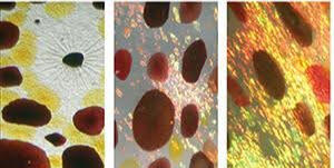
Chromatophores are pigment-containing or light-reflecting organs in amphibians, reptiles, fish, crustaceans, and cephalopods.
Color changes are the result of specialized cell structures interacting with ambient light from the environment.Chromatophore Cell Types:
Chromatophores Pigmented - Melanophores (black/brown), Xanophores (yellow), Erythrophores (red/orange)
Iridophores Reflective
Leucophores Reflective
Together, these cells can create any color in the visible spectrum.
Layers Layers
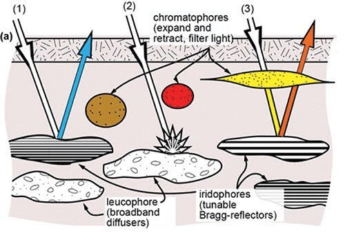
Each cell type uses a different mechanism to manipulate light.
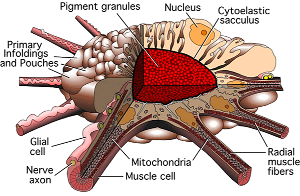
As this cell expands, the pigment inside becomes visible.
Each chromatophore contains Yellow, Red, or Brown type Pigment.Combined with multiple layers of reflectors to enable the animal to reflect any wavelength of visible light.
Pigment-containing chromatophores have around 20 radial muscle fibers. These cells unfold fast (milliseconds).
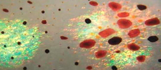
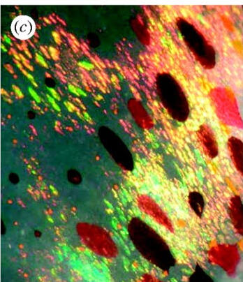
Chromatophores & iridophores work together to create optical diversity
Red and yellow chromatophores transmit much of the incoming light and play an important role in modulating iridescence.
Brown chromatophores transmit less light and so are used to create dark patches or block iridescence/ polarized light.
Iridophores – multilayer reflectors
Thin plates that reflect light by thin film interference (Bragg Reflector).The more oblique the angle of incidence, the shorter the peak wavelength of the light reflected.
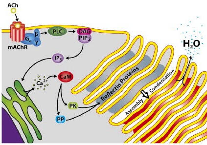
Increasing acetylcholine concentration causes the cells to stack more tightly which alters the reflectance of the iridophore.
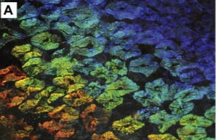
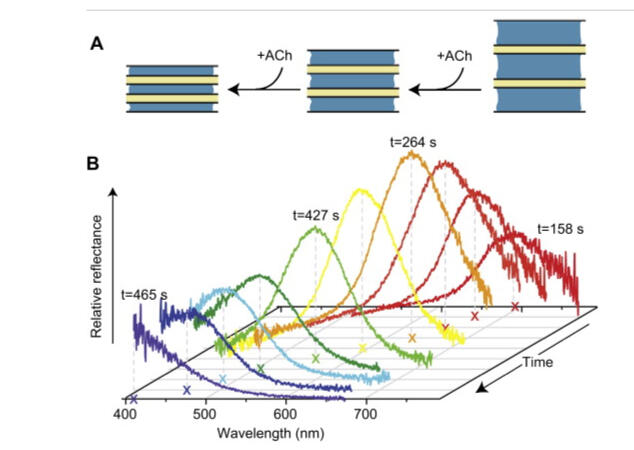
Multiple Bragg reflectors work together to create an overall emission depending on the angle of incidence.
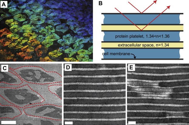
Reflective structures are made up of proteins called Reflectin.Reflectin changes its density upon condensation and the cells use this to turn on/off iridophores.
Iridophores
The membrane invaginations provide a high surface area interface between all of the protein lamellae and the extracellular space.The morphology provides a route for the rapid exchange of water facilitated by the large surface area between the protein lamellae and extracellular space.

Leucophores – (white)
Leucophores contain spherical reflectin protein assemblages that scatter light equally well in all directions (ie, diffusion / white light)
They provide a backdrop against which chromatophores (and iridophores) can create high contrast patterns
Leucophores reflect the ambient spectrum (they look red in red light, blue in blue light, etc.)
References
Dinneen S.R., Osgood R.M., J. Phys. Chem. A., 2017, 8, 313-317
Mäthger L.M., Hanlon R.T., Cell Tissue Res., 2007, 329, 179–186
DeMartini D.G., Krogstad D.V., Morse D.E., PNAS., 2013, 110, 2552-2556
Ghoshal A., DeMartini D.G., Eck E., Morse D.E., J R Soc., 2013, 10
Tao A.R., DeMartini D.G., Biomaterials, 2010, 31, 793-801
Cooper K.M., Hanlon R.T., Budelmann B.U., Cell Tissue Res., 1990, 259, 15-24
Phan L., Kautz R., Leung E.M., Naughton K.L., Van Dyke Y., Gorodetsky A.A., Chem. Mater., 2016, 28, pp 6804–6816
Cloney R. A., E. Florey., Zellforsch, 1968, 89, 250‐280
Mäthger L.M., Denton E.J., Marshall J.N., Hanlon R.T., J R Soc, 2008, 6, 149-163
Mäthger L.M., Denton E.J., Marshall J.N., Hanlon R.T., J R Soc, 2008, 6, 149-163
Messenger J. B., Biol. Rev., 2001, 76, 473–528
Kingston A.C. N., Kuzirian A.M., Hanlon R.T., Cronin T.W., J. Exp. Biol., 2015, 218, 1596-1602